The Science of Recovery: How Your Body Heals Tendons, Ligaments, and Muscle — And What Peptides May Add
Tendons and ligaments heal slowly — and often incompletely. Here's the biology behind why, and what the research on BPC-157, TB-500, and GHK-Cu says about where peptides might fit in the recovery process.
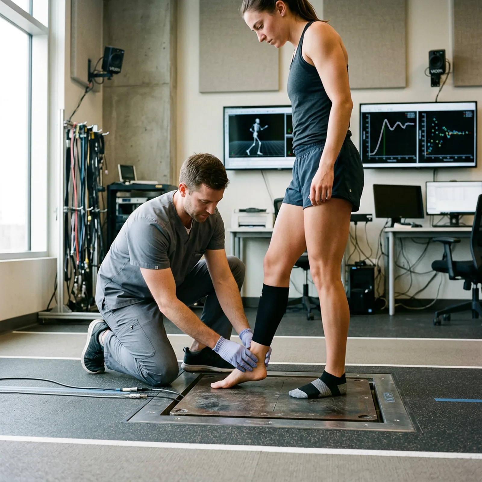
Why Injuries to Tendons and Ligaments Are So Frustrating
You've probably noticed that a muscle strain heals in days. A ligament tear? Months. And a tendon rupture might leave you dealing with compromised tissue for years — even after it's technically "healed." That disparity isn't bad luck. There's a clear biological reason for it, and understanding it changes how you think about recovery research.
Muscles are vascular tissue. They're packed with blood vessels that deliver oxygen, growth factors, and repair cells almost immediately after injury. Tendons and ligaments are a different story. They're largely avascular — meaning blood supply is sparse, particularly in the central regions where tears most often occur. Less blood flow means slower delivery of everything your body needs to rebuild.
Sharma and Maffulli laid out the cellular and mechanical architecture of this problem clearly in a 2006 paper in the Journal of Musculoskeletal and Neuronal Interactions. Their analysis showed that despite multiple overlapping repair phases, "the biochemical and mechanical properties of healed tendon tissue never match those of intact tendon." That's not a pessimistic framing — it's a scientific baseline. And it's exactly the kind of gap that peptide researchers have started trying to address.
The Three Phases of Connective Tissue Healing
Tendon and ligament repair follows a predictable sequence that unfolds over weeks to months. Understanding this sequence matters because different interventions — including peptides — may operate differently depending on which phase is active.
Phase 1: Inflammation (Days 1–7)
Immediately after injury, erythrocytes and inflammatory cells flood the site. Within the first 24 hours, monocytes and macrophages take over, clearing debris through phagocytosis. Vasoactive mediators cause swelling. Blood vessels dilate. The whole process feels miserable — and it's supposed to. Inflammation is how your body signals the repair machinery to mobilize.
Here's where it gets interesting, though. Chronic inflammation — the kind that lingers past the first week — starts working against you. Prolonged exposure to inflammatory cytokines can impair fibroblast function and degrade newly forming collagen. So the challenge isn't suppressing inflammation outright. It's timing.
Phase 2: Proliferation (Weeks 2–6)
This is when the real construction work begins. Tenocytes and fibroblasts migrate into the wound bed, proliferate, and begin synthesizing collagen. Early collagen is predominantly Type III — weaker, more disorganized. Over time it shifts toward the mechanically superior Type I. The extracellular matrix (ECM) starts to fill in, blood vessel growth occurs (angiogenesis), and the structural scaffolding of new tissue takes shape.
Cell migration is rate-limiting here. How fast fibroblasts can reach the injury site — and how well they survive once they arrive — determines how quickly and completely the proliferative phase proceeds. This is relevant to peptide research because several compounds have been studied specifically for their effects on fibroblast migration speed and stress survival.
Phase 3: Remodeling (Months 3–18+)
Once the initial scaffold is down, the tissue spends months reorganizing. Collagen fibers align along mechanical stress lines. Matrix metalloproteinases (MMPs) degrade excess or disorganized matrix while new fibers are laid in. Even after the tissue appears macroscopically healed, remodeling continues. This is why "healed" tendons remain vulnerable to reinjury: they've closed, but the internal architecture hasn't fully matured.
Where Peptide Research Fits In
The peptides most studied for connective tissue repair fall into a few mechanistic categories. Some appear to accelerate fibroblast activity. Others promote angiogenesis. Some modulate the inflammatory response. A few may do all three to varying degrees, which is part of why they attract research interest in the first place.
BPC-157: The Fibroblast Migration Story
BPC-157 is a 15-amino-acid peptide originally isolated from human gastric juice. Most of its early research focused on gastrointestinal tissue — ulcers, fistulas, colon healing. But researchers in Taiwan noticed that the same angiogenic and fibroblast-activating mechanisms might apply to musculoskeletal tissue, and they started testing it in tendon models.
The key study here comes from Chang et al., published in the Journal of Applied Physiology in 2011. Their group isolated Achilles tendon fibroblasts from rats and exposed them to BPC-157 at various concentrations. The results: BPC-157 significantly accelerated the outgrowth of tendon fibroblasts from explant tissue. More striking was the migration finding — treated cells migrated 2.3 times faster than controls in transwell assays. The mechanism appears to involve activation of the FAK-paxillin signaling pathway, which governs how cells attach to and move through extracellular matrix. BPC-157 also enhanced cell survival under oxidative stress (the H₂O₂ model), which matters because the healing wound environment is oxidatively demanding.
A 2014 follow-up study expanded on this, showing that BPC-157 increases growth hormone receptor expression in tendon fibroblasts — effectively sensitizing them to GH's proliferative signals. This suggests BPC-157 may not just act directly, but also amplify the tissue's responsiveness to other healing signals already in circulation. We found this particularly interesting because it implies BPC-157 could act synergistically with the body's own repair mechanisms rather than replacing them.
In animal ligament models, BPC-157 consistently improved outcomes — better functional recovery, stronger tissue on load-to-failure testing, and histologically cleaner collagen architecture. It's also been studied in the context of corticosteroid-impaired healing, where it appears to counteract some of the tendon-weakening effects steroids can have. Worth noting: a 2025 review in an orthopedic journal summarizing 36 studies from 1993–2024 found consistent preclinical support for BPC-157 in musculoskeletal injury models. Clinical human data remains limited, though a small retrospective study reported pain relief in 7 of 12 patients with chronic knee pain following intraarticular injection.
For researchers interested in BPC-157, 22EXO's BPC-157 (5mg) is available in research-grade lyophilized form.
TB-500: The Actin Angle
TB-500 is the synthetic peptide fragment corresponding to amino acids 17–23 of thymosin beta-4 (Tβ4), a naturally occurring protein found in virtually every human cell. The specific sequence — LKKTET — is the region responsible for actin binding, and actin regulation is central to cell migration. Without actin polymerization and depolymerization, cells can't move. And if cells can't move, wounds don't close.
Thymosin beta-4 research has shown that this protein promotes the mobilization, migration, and differentiation of stem and progenitor cells that form new blood vessels and regenerate tissue. It also decreases the density of myofibroblasts in healing wounds — a meaningful finding because excess myofibroblasts are the cellular driver of fibrosis and scar formation. Less scar, more functional tissue.
In dermal Phase II clinical trials, thymosin beta-4 accelerated wound healing and was well tolerated in patients with pressure ulcers, stasis ulcers, and epidermolysis bullosa. TB-500 mirrors the active region of this molecule. Animal models have shown benefits in bone, tendon, and cardiac healing. Human data is more limited.
22EXO's TB-500 (5mg) is available for research purposes.
GHK-Cu: Collagen Architecture and the Anti-Inflammatory Angle
Copper peptide GHK-Cu (glycine-histidine-lysine complexed with copper) takes a different approach to connective tissue repair. Rather than focusing specifically on fibroblast migration, GHK-Cu appears to modulate both collagen synthesis and breakdown simultaneously — acting as a regulator rather than simply a stimulant.
Research has shown GHK-Cu at nanomolar concentrations stimulates both synthesis and breakdown of collagen and glycosaminoglycans, modulates MMP activity (the enzymes responsible for ECM remodeling), and reduces pro-inflammatory cytokines including TNF-alpha and IL-6. In wound models ranging from rabbit skin to ischemic rat tissue, GHK-Cu consistently improved healing speed, collagen organization, and antioxidant enzyme activity.
Its role in recovery research is more as a modulator of the remodeling phase than an acute repair accelerant — though the two aren't cleanly separable in practice. GHK-Cu (50mg) is available from 22EXO for research applications.
The Case for Combination Research Protocols
Because BPC-157 and TB-500 operate through different mechanisms — FAK-paxillin signaling and actin regulation, respectively — researchers have begun studying them together. The logic: if one enhances fibroblast migration and oxidative stress tolerance, and the other promotes angiogenesis and stem cell recruitment, combining them might address more of the healing cascade simultaneously.
There isn't yet published human clinical data comparing combination protocols to single-peptide approaches. What exists is mechanistic rationale plus the absence of known antagonistic interactions. Some preclinical work suggests additive or complementary effects, but that finding should be treated with appropriate epistemic caution.
For researchers interested in exploring this angle, 22EXO's BPC-157/TB-500 Blend (5mg) combines both peptides in a single lyophilized preparation.
What the Research Doesn't Yet Tell Us
The honest picture: nearly all the compelling evidence for BPC-157 and TB-500 in connective tissue comes from animal models — mostly rats. Controlled human clinical trials are sparse. Dosing, timing relative to injury phase, and route of administration remain poorly characterized in human subjects. The 2025 orthopedic review was candid about this: preclinical results are consistently positive, but there is "no clinical safety data in humans" for BPC-157 beyond case series.
This doesn't invalidate the research direction — animal connective tissue models are genuinely informative about biology. But it does mean researchers should hold their conclusions appropriately. The mechanistic story is plausible and internally consistent. Whether it translates cleanly to human clinical outcomes, and at what doses, remains an open question worth investigating.
Practical Considerations for the Research-Minded
A few things worth understanding if you're evaluating connective tissue healing peptides:
- Phase specificity matters. Interventions that help during the proliferative phase may behave differently during late remodeling. Timing of administration relative to injury timeline is an under-studied variable in most existing research.
- Purity is not trivial. Peptide research compounds vary enormously in actual purity and sequence fidelity. Third-party HPLC testing is the minimum standard for trustworthy research supplies.
- The body also heals on its own. Any research protocol should account for the natural time course. Connective tissue outcomes at 6 months look very different from outcomes at 6 weeks regardless of intervention.
For more on how BPC-157 compares mechanistically to TB-500, see our BPC-157 vs. TB-500 Research Comparison. Our piece on peptide blends for healing research explores the combination rationale in more depth, and if you want the mechanism detail on BPC-157 specifically, we've written a dedicated piece on how BPC-157 works.
Frequently Asked Questions
Why do tendons heal more slowly than muscles?
Tendons are largely avascular — they have poor blood supply, particularly in their central regions. Blood is what delivers repair cells, growth factors, and oxygen to injured tissue. With less of it flowing through tendons, every stage of the repair process takes longer than in highly vascular muscle tissue.
What is the FAK-paxillin pathway and why does it matter for healing?
FAK (focal adhesion kinase) and paxillin are proteins involved in how cells attach to and move through extracellular matrix. When this pathway is activated, fibroblasts migrate faster to injury sites — which is critical because cell migration is one of the rate-limiting steps in tendon repair. Chang et al. (2011) showed <a href="/product/bpc-157-5mg">BPC-157</a> activates this pathway in tendon fibroblasts.
What is the difference between BPC-157 and TB-500 mechanistically?
<a href="/product/bpc-157-5mg">BPC-157</a> primarily acts on fibroblast migration and survival via the FAK-paxillin pathway, and may also increase growth hormone receptor sensitivity in tendon cells. <a href="/product/tb-500-5mg">TB-500</a> (a fragment of thymosin beta-4) works primarily through actin regulation — it binds actin and promotes cell migration, angiogenesis, and stem cell recruitment. The mechanisms are complementary rather than redundant.
Are these peptides approved for human use?
No. <a href="/product/bpc-157-5mg">BPC-157</a>, TB-500, and <a href="/product/ghk-cu-50mg">GHK-Cu</a> are research compounds. They are not FDA-approved for human therapeutic use. All of 22EXO's peptides are sold strictly for research purposes and are not intended for human consumption.
What does the collagen remodeling phase involve?
During remodeling (which begins around 3 months post-injury and can continue for 18+ months), collagen fibers align along mechanical stress lines, matrix metalloproteinases degrade disorganized matrix, and Type III collagen gradually transitions to stronger Type I collagen. Even tissue that appears healed externally may still be undergoing internal architectural reorganization.