How to Reconstitute Peptides: Step-by-Step Lab Protocol
Lyophilized peptides arrive as dry powder or cake. Research-chemical reconstitution requires the right solvent, the slow-stream technique, and concentration math to keep a research solution intact.
How to Reconstitute Peptides: Step-by-Step Lab Protocol
What Reconstitution Means in a Research Context
Lyophilization — freeze-drying — removes water from a peptide solution by sublimation under vacuum. The vial you receive contains a dry solid: the peptide plus any excipients used during the freeze-drying cycle, locked in the spatial arrangement they held in solution. Reconstitution reverses that process: add solvent, dissolve the solid, and produce an aqueous research solution suitable for in-vitro assays, cell-culture work, or other non-clinical laboratory applications.
Getting this right requires attention to three variables: which solvent you use, how you introduce it to the vial, and how you handle and store the resulting research solution. Each step has a documented failure mode that degrades or destroys the research chemical before a single assay is run.
Step 1 — Vial Inspection Before Research-Chemical Reconstitution
Before handling the solvent or syringe, hold the vial up to a bench light and examine the lyophilized material. A properly lyophilized peptide produces a coherent, white to off-white cake at the bottom of the vial. The cake holds its shape when the vial is tilted slowly. Collapsed powder, dark discoloration (yellowing, browning), or visible moisture condensation on the interior glass wall are all signs of temperature excursion during shipping or storage — conditions that may have compromised the research chemical before your lab received it.
Check the stopper separately. The rubber septum must be seated flush and uniformly around its perimeter. A partially dislodged stopper introduces contamination risk that no downstream sterile technique can fully correct. Every vial supplied with a Certificate of Analysis (COA) and HPLC ≥98% documentation should show consistent cake morphology and seating lot-to-lot. If either looks abnormal, do not proceed — contact the supplier with a photograph of the vial before reconstitution.
Step 2 — Solvent Selection for Peptide Research Solutions
For the large majority of lyophilized research peptides, bacteriostatic water (sterile water for injection, USP, containing 0.9% benzyl alcohol) is the appropriate solvent when the reconstituted research solution will be used across multiple draws over days or weeks. The benzyl alcohol is a bacteriostatic and fungistatic preservative that inhibits microbial growth after the vial stopper has been punctured, extending the working window of the research solution to approximately 28 days at 2–8°C.
Plain sterile water for injection carries no antimicrobial protection. Once punctured, it is effectively single-use; the entire volume should be consumed within 24 hours or discarded. For research protocols requiring only a single aliquot preparation, sterile water is acceptable. For any multi-draw workflow — which describes most routine lab protocols — bacteriostatic water is the standard.
A small number of research peptides dissolve poorly in neutral aqueous solution at room temperature. The standard lab workaround is to first add a small volume of 0.1–1% acetic acid in sterile water to begin dissolution, then bring the volume to target with bacteriostatic water. Some protocols use 5–10% DMSO as a co-solvent for hydrophobic sequences, though this introduces variables in downstream cell-culture assays and should only be used when aqueous dissolution genuinely fails after 15 minutes of gentle inversion.
For two of the most widely studied tissue-remodeling research peptides — BPC-157 (5 mg, CAS 137525-51-0) and TB-500 (5 mg, CAS 77591-33-4) — plain bacteriostatic water at room temperature dissolves both readily without co-solvent. Neither requires acidic pre-treatment under standard lab conditions.
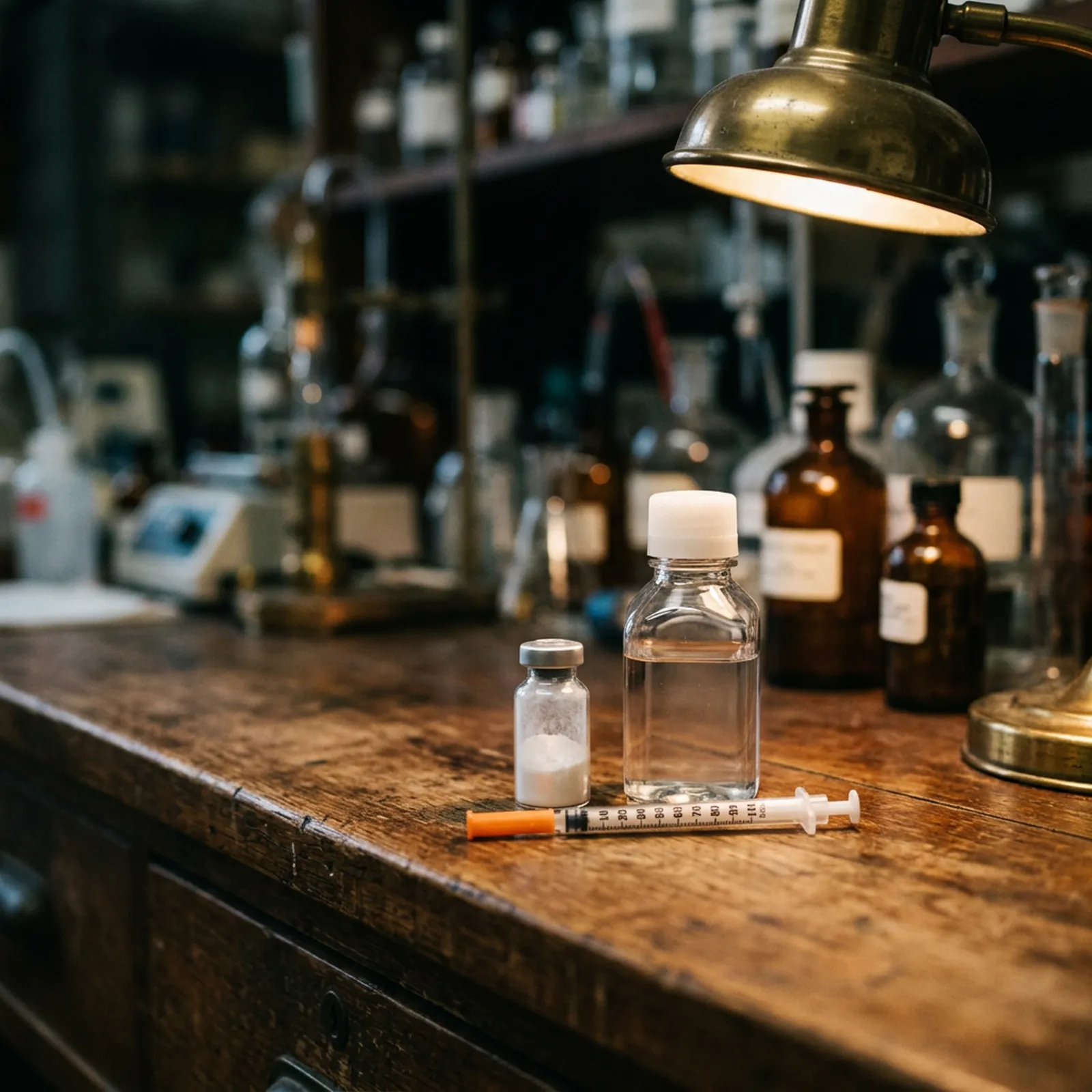
Step 3 — The Slow-Stream Technique
How you add solvent to the lyophilized cake is as important as which solvent you choose. The correct technique in a research lab setting:
- Draw your calculated volume of bacteriostatic water into a sterile syringe (see concentration math below).
- Wipe the vial stopper with a 70% isopropanol swab. Allow it to dry for 15–20 seconds before inserting the needle — wet alcohol on the needle tip is a contamination route into the vial.
- Insert the needle at an angle with the bevel facing up, directing the needle tip toward the inner glass wall of the vial rather than pointing at the cake.
- Depress the plunger slowly, directing the solvent stream down the glass wall so it flows over the lyophilized cake gradually.
- Remove the syringe and set the vial aside without agitation.
Directing the stream toward the glass wall avoids mechanical disruption of the lyophilized matrix. A direct stream onto the cake shears the peptide structure and introduces air bubbles that expand the interfacial surface area where peptide denaturation occurs.
Step 4 — Dissolution: Gentle Inversion Only
After adding solvent, allow the vial to sit at room temperature for 2–5 minutes. Most research peptides dissolve passively in this time. If the research solution remains cloudy or partially undissolved after 5 minutes, gently roll the vial between your fingers or slowly invert it 3–5 times and wait another 5 minutes.
Do not vortex. Do not shake. Both generate turbulent flow and a high density of air-water interfaces. Peptide molecules adsorb to those interfaces and can aggregate or form insoluble particulates. For a 43-residue peptide like TB-500 — which has amphiphilic secondary structure — shaking is the fastest way to reduce the biologically active fraction in your research solution without producing any visible evidence of the loss. The solution may appear clear post-shaking while containing substantially fewer intact molecules than before.
TB-500 specifically may take 10–15 minutes to fully dissolve; larger peptides are slower than small ones. Patience at this stage is part of the protocol, not an optional detail.
Step 5 — Concentration Math for Research Dilution Preparation
The dilution relationship is: Concentration (mg/mL) = Mass (mg) ÷ Volume added (mL).
To work backward from a target stock concentration: Volume to add (mL) = Mass (mg) ÷ Target concentration (mg/mL).
Common research stock setups from a 5 mg vial:
- + 5 mL BAC water → 1 mg/mL (1,000 mcg/mL) stock
- + 2.5 mL BAC water → 2 mg/mL (2,000 mcg/mL) stock
- + 10 mL BAC water → 0.5 mg/mL (500 mcg/mL) stock
Once the stock concentration is established, calculating the volume for a specific research sample aliquot is straightforward division. If your stock is 1 mg/mL and your protocol calls for a 250 mcg sample, the draw volume is 0.25 mL (250 mcg ÷ 1,000 mcg/mL). On a U-100 insulin syringe, that corresponds to the 25-unit mark. Label every vial with its stock concentration and the date of reconstitution at the time of preparation.
Reconstitution Calculator
For research lab dilution math only. Not for human use.
Step 6 — Storage of Reconstituted Research Solutions
Reconstituted research solutions should be transferred to 2–8°C storage immediately after preparation. Use a stable shelf position — not the refrigerator door, where temperature swings with each opening, and not against the back wall if your unit freezes there. Protect from light exposure; amber vials are preferable, or wrap clear vials in aluminum foil.
Stability windows for research peptide solutions in bacteriostatic water at 2–8°C:
- 14–28 days: standard guidance for most peptides. BPC-157 (CAS 137525-51-0, the 15-residue sequence GEPPGKPADDAGLV) maintains documented activity across a 28-day refrigerated window under proper storage. TB-500 (Ac-LKKTETQ fragment) shows comparable stability.
- ≤14 days: recommended for peptides containing methionine residues or disulfide bonds, which are more susceptible to oxidative degradation in aqueous solution.
Three Practices That Destroy Reconstituted Research Solutions
Freezing the reconstituted vial. Once bacteriostatic water has been added, do not freeze the vial to attempt extended storage. Ice crystal formation mechanically disrupts peptide structure, and the expansion-contraction cycle promotes aggregation. Lyophilized powder stores well at −20°C for months; reconstituted research solutions do not. Prepare only the volume your protocol requires for the current study window.
Contaminating the stopper. Every needle puncture carries a contamination risk. Wipe with isopropanol before each entry into the vial — without exception. After 30–40 punctures, the mechanical integrity of standard butyl rubber stoppers degrades, and micro-coring (small rubber fragments entering the vial) becomes a real risk. For high-throughput labs drawing from the same vial multiple times per day, tracking puncture count is practical quality control.
Temperature cycling the vial. Removing a reconstituted vial from refrigeration, leaving it at bench temperature for an extended protocol session, then returning it — repeated across multiple days — accelerates peptide degradation. The standard lab practice is to aliquot the volume needed for a given session before the main vial leaves the refrigerator. Discard any aliquot not used in the session rather than returning it to the stock vial.
Peptide-Specific Notes
BPC-157 Research Solutions
BPC-157 is a 15-amino-acid synthetic peptide (pentadecapeptide, MW 1419.5 Da) derived from a partial sequence of human body protection compound. It dissolves readily in bacteriostatic water at room temperature within 2–5 minutes of gentle inversion. Research stock concentrations of 500 mcg/mL to 1 mg/mL are most common in published in-vivo rodent protocols (typically 10–100 mcg/kg ranges), which sets the practical context for dilution math in those experimental designs. All purity specifications — HPLC ≥98%, COA on every batch — apply to the lyophilized material at time of manufacture; handling and storage after reconstitution are the responsibility of the receiving laboratory.
TB-500 Research Solutions
TB-500 (thymosin beta-4 C-terminal fragment, 43 amino acids, MW ~4963 Da) dissolves more slowly than most sub-20-residue peptides. Plan for 10–15 minutes of passive dissolution after adding bacteriostatic water before concluding there is a solubility problem. The peptide is acutely sensitive to the no-shaking rule: its length and amphiphilic character make it among the most aggregation-prone peptides in common research use. Confirm complete dissolution visually (clear to very slightly opalescent solution) before drawing any research sample.
Blend Vials
Pre-blended vials — such as the BPC-157 + TB-500 combination formats — follow an identical reconstitution protocol. The mass listed on the label is the combined peptide mass. Check the product COA for the per-component mass ratio if your protocol requires per-peptide concentration calculations.
Final Quality Check
After full dissolution, the research solution should be clear to very slightly opalescent, colorless unless the peptide itself has characteristic color (GHK-Cu solutions are characteristically blue, for example). Any visible particulates, persistent cloudiness after 15 minutes of inversion, or unexpected color indicate a problem. A COA confirming HPLC ≥98% at the lyophilized stage does not guarantee that reconstitution errors have not degraded the material — visual inspection of the final research solution is the last quality gate under the lab's control.
All products supplied by 22EXO are Research Use Only (RUO) compounds. They are not intended for human or veterinary administration. This protocol describes research-chemical reconstitution for in-vitro, cell-culture, and non-clinical laboratory applications only.
Frequently Asked Questions
What solvent should a research lab use to reconstitute peptides?
Bacteriostatic water (sterile water for injection, USP, containing 0.9% benzyl alcohol) is the standard solvent for most lyophilized research peptides when the reconstituted research solution will be drawn across multiple sessions. The benzyl alcohol preservative inhibits microbial growth after the stopper is punctured, extending the stability window of the research solution to approximately 28 days at 2–8°C. For single-session preparations where the entire volume will be used within 24 hours, plain sterile water for injection is acceptable. Some hydrophobic peptide sequences require a dilute acetic acid pre-treatment or a small percentage of DMSO as co-solvent before dilution in bacteriostatic water.
How do you calculate how much BAC water to add for a target stock concentration?
The formula is: volume to add (mL) = vial mass (mg) ÷ target concentration (mg/mL). For a 5 mg vial where you want a 1 mg/mL research stock, add 5 mL of bacteriostatic water. For 2 mg/mL, add 2.5 mL. For 0.5 mg/mL, add 10 mL. Once the stock is prepared, the volume for a specific research sample aliquot is target mass (mcg) ÷ stock concentration (mcg/mL). The embedded calculator in this article automates this math for standard vial and syringe combinations.
How long is a reconstituted peptide research solution stable at 2–8°C?
Most research peptide solutions in bacteriostatic water remain stable for 14–28 days at 2–8°C when stored away from light and temperature fluctuation. <a href="/product/bpc-157-5mg">BPC-157</a> (CAS 137525-51-0) and <a href="/product/tb-500-5mg">TB-500</a> (CAS 77591-33-4) are documented at approximately 28 days under proper refrigerated storage conditions. Peptides containing methionine residues or free disulfide bonds are more susceptible to oxidative degradation and should be used within 14 days. Freeze-thaw cycling of reconstituted solutions accelerates degradation and should be avoided.
Why is shaking a reconstituted peptide vial harmful?
Rapid mechanical agitation from shaking generates high-energy air-water interfaces at a rate far exceeding what occurs in gentle inversion. Peptide molecules adsorb preferentially to those interfaces and can misfold, aggregate, or form insoluble particulates. This effect is particularly pronounced for larger polypeptides like <a href="/product/tb-500-5mg">TB-500</a> (43 residues, partial helical secondary structure) and any peptide with amphiphilic character. The active content of a shaken research solution may be measurably lower than before shaking even though the solution appears clear, because aggregated peptide is not visible to the naked eye at typical research concentrations.
What does the appearance of the lyophilized cake indicate about product quality?
A properly lyophilized research peptide forms a coherent, white to off-white cake that fills the vial base and holds its shape when tilted. Collapsed powder, discoloration (yellowing or browning), or moisture condensation on the inner glass wall indicate temperature excursion during lyophilization, shipping, or storage — all of which can compromise the research chemical prior to <a href="/blog/peptide-reconstitution-handling-guide">reconstitution</a>. Every lot supplied with <a href="/blog/peptide-purity-hplc-testing-guide">HPLC</a> ≥98% certification and a batch COA should show consistent cake morphology. Abnormal cake appearance warrants contact with the supplier before reconstitution proceeds.