Polypeptide vs Peptide vs Protein: Definitions, Sizes, and Why It Matters
The terms peptide, polypeptide, and protein are used interchangeably in casual conversation and inconsistently in the primary literature. The distinctions are real, biologically meaningful, and directly relevant to how researchers synthesize, store, and handle these molecules in the lab.
Why These Terms Keep Getting Conflated
In a molecular biology lecture, an undergraduate learns that proteins are large polypeptides. In a peptide chemistry paper, a 43-residue molecule gets called a polypeptide in the abstract and a peptide in the methods. In a pharmacology textbook, insulin is described as a peptide hormone despite having 51 amino acids — above most textbook cutoffs for the term 'peptide.' The confusion is not random. It reflects genuine ambiguity at the boundaries between these categories, compounded by the fact that different scientific subfields have different conventions for where the lines sit.
This article uses the biochemical definitions most commonly applied in research settings, explains where the conventional thresholds come from, identifies where they break down, and draws out the practical implications for synthesis, storage, and experimental design. Understanding these distinctions is directly useful for any lab working with synthetic RUO peptides such as BPC-157 or TB-500.
Amino Acids: The Shared Building Block
Every peptide, polypeptide, and protein is assembled from the same 20 standard alpha-amino acids (plus the two less common coded residues selenocysteine and pyrrolysine). Each amino acid consists of a central carbon atom (the alpha-carbon) bearing four substituents: an amino group (–NH2), a carboxyl group (–COOH), a hydrogen atom, and a variable side chain (R group) that determines the amino acid's chemical character. Glycine has R = H, making it the smallest and the only achiral standard amino acid. Tryptophan at R = an indole group is the largest. The 20 standard side chains span a range from charged (aspartate, glutamate, lysine, arginine, histidine) to polar neutral (serine, threonine, asparagine, glutamine, cysteine, tyrosine) to nonpolar aliphatic (glycine, alanine, valine, leucine, isoleucine, methionine, proline) to aromatic (phenylalanine, tyrosine, tryptophan).
When two amino acids react, the carboxyl group of one condenses with the amino group of the other, releasing a water molecule and forming a covalent amide linkage called a peptide bond. The product is a dipeptide. Add a third amino acid and you have a tripeptide. Continue the process and you build a chain — a sequence defined by the identity of each residue and the order in which they appear. That sequence is the primary structure.
Definitions and Conventional Size Thresholds
The three terms — peptide, polypeptide, protein — describe regions along a continuous spectrum of chain length, and the boundaries between them are conventional rather than absolute.
Peptide is broadly applied to chains containing up to approximately 50 amino acid residues, though many sources set the upper boundary at 20–30 residues. The IUPAC definition does not impose a length restriction on 'peptide' at all — technically any chain of amino acids joined by peptide bonds qualifies. In practice, researchers call short chains (2–20 residues) peptides without qualification. At the lower end: dipeptides (2 residues), tripeptides (3 residues), and the oligopeptide designation (2–20 residues). BPC-157 at 15 residues (sequence: Gly-Glu-Pro-Pro-Pro-Gly-Lys-Pro-Ala-Asp-Asp-Ala-Gly-Leu-Val, CAS 137525-51-0) is unambiguously a peptide by any definition.
Polypeptide in the strictest chemical sense means any chain of amino acids — 'poly' simply means 'many.' But in practice, the term is reserved for the intermediate zone between short peptides and folded proteins, roughly 20 to 50+ residues. TB-500 at 43 residues (CAS 77591-33-4, the primary bioactive sequence of thymosin beta-4, acetylated at the N-terminus: Ac-Ser-Asp-Lys-Pro-Asp-Met-Ala-Glu-Ile-Glu-Lys-Phe-Asp-Lys-Ser-Lys-Leu-Lys-Lys-Thr) sits squarely in polypeptide territory by most conventions. It is too long to be called a short peptide without qualification, too short to be confidently called a protein, and it does not adopt a stable globular fold in solution.
Protein is generally applied to chains of more than 50 amino acid residues that fold into a defined three-dimensional structure. The fold is what functionally distinguishes proteins from longer polypeptides: a random-coil 60-residue chain is not, in most practical usages, a protein. Insulin (51 total residues, A-chain of 21 aa and B-chain of 30 aa connected by two interchain disulfide bonds) occupies a boundary position — it is consistently called a protein in endocrinology literature and a 'peptide hormone' in other contexts. The average single-domain protein is approximately 200–400 residues. Titin, the largest known protein in the human body, contains approximately 34,350 residues and has a molecular weight of around 3,800 kDa.
Where the Line Between Peptide and Protein Gets Blurry
The insulin case illustrates the core problem: size alone is not sufficient to classify a molecule. Insulin at 51 residues adopts a well-defined tertiary structure (a compact globular fold stabilized by three disulfide bonds), circulates in blood at nanomolar concentrations, and has a clinically defined receptor-binding mechanism. It is functionally a protein. At the same time, it is smaller than many peptides synthesized by SPPS for research purposes, and it was historically studied within the tradition of peptide hormone biology. The term 'peptide hormone' has stuck in pharmacology for molecules like insulin, glucagon (29 residues), ACTH (39 residues), and GnRH (10 residues) — none of which are peptides by any structural definition that requires absence of tertiary structure.
Conversely, some synthetic polypeptides of 80–100 residues produced by SPPS are called peptides in the synthetic chemistry literature simply because they were made by chemical synthesis rather than recombinant expression. The naming reflects the synthetic tradition more than the molecule's biophysical properties.
The most defensible distinction is structural: a protein is a polypeptide chain that adopts a stable, specific three-dimensional fold under physiological conditions. A peptide lacks this stable fold — it may have local conformational preferences or exist in solution as a distribution of conformers, but it does not have a single dominant tertiary structure. This structural definition maps onto function better than any simple amino acid count, and it also maps onto the practical handling differences described in the final section of this article.
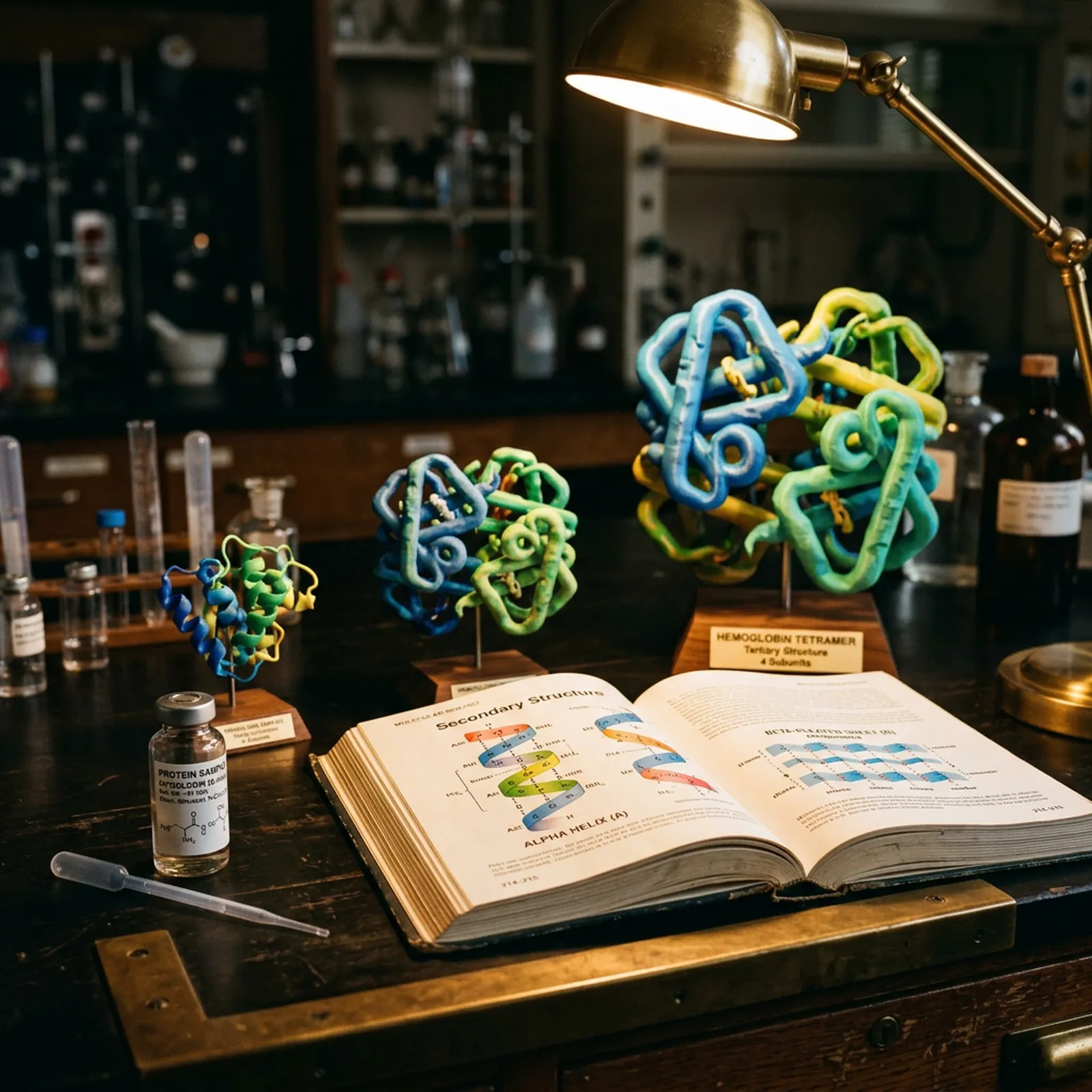
Structural Hierarchy: From Primary to Quaternary
Regardless of size, the structural organization of polypeptides and proteins is described using four levels.
Primary structure is the linear sequence of amino acids. It is the foundational information from which all higher-order structure derives. For BPC-157, the 15-residue sequence (Gly-Glu-Pro-Pro-Pro-Gly-Lys-Pro-Ala-Asp-Asp-Ala-Gly-Leu-Val) is sufficient to fully specify the molecule — there are no disulfide bonds or post-translational modifications in the synthetic version.
Secondary structure refers to local, regular patterns of hydrogen bonding between backbone atoms. The two dominant elements are the alpha-helix, a right-handed helical arrangement stabilized by H-bonds between the carbonyl oxygen of residue i and the amide hydrogen of residue i+4, and the beta-sheet, stabilized by H-bonds between adjacent extended strands running parallel or antiparallel to each other. A third important element is the beta-turn or type-I/II beta-turn, a four-residue structure that reverses chain direction and is particularly common in short bioactive peptides. Proline residues — which BPC-157 contains three of in its Pro-Pro-Pro motif at positions 3–5 — are known as secondary structure breakers because the nitrogen of proline is incorporated into its pyrrolidine ring and cannot donate an amide hydrogen bond, which disrupts helical structures. The poly-proline stretch in BPC-157 confers a locally extended, semi-rigid conformation that may be relevant to its observed receptor interactions in preclinical models.
Thymosin beta-4, the full-length precursor of TB-500's bioactive sequence, contains a central alpha-helical domain (approximately residues 17–30 in the full 44-residue protein) that mediates G-actin binding. This helix is preserved in the TB-500 sequence and represents one of the few cases in which a short polypeptide of this size has a documented, functionally relevant secondary structural element.
Tertiary structure is the overall three-dimensional arrangement of a single polypeptide chain, including all its secondary structure elements and the loops connecting them. Tertiary structure is stabilized by the cumulative effect of hydrophobic packing (nonpolar side chains buried in the protein core), disulfide bonds (covalent S–S linkages between cysteine residues), electrostatic interactions between charged side chains, and hydrogen bonding beyond the backbone H-bonds of secondary structure. Short peptides and most polypeptides below roughly 50 residues do not form stable tertiary structures in solution under most conditions. Their biological activity depends on specific short sequence motifs that adopt defined conformations transiently upon receptor binding rather than in the free solution state.
Quaternary structure applies only to proteins with more than one polypeptide chain (subunit). Hemoglobin (2 alpha + 2 beta subunits), collagen (triple helix of three alpha chains), and many enzyme complexes are examples. No peptide or short polypeptide in the research context covered by this site exhibits quaternary structure.
Synthesis Differences: SPPS vs. Recombinant Protein Expression
The production method for a molecule is directly tied to its size, and understanding the difference helps researchers evaluate the quality and limitations of what they are purchasing.
Solid-phase peptide synthesis (SPPS), introduced by Merrifield in 1963, is the standard method for producing synthetic peptides and polypeptides up to approximately 50 residues. In Fmoc SPPS (the modern standard, using 9-fluorenylmethyloxycarbonyl temporary protecting groups), the peptide chain is built C-terminus to N-terminus on a polystyrene-based resin. Each cycle involves: deprotection of the terminal Fmoc group with 20% piperidine in DMF, washing, coupling of the next Fmoc-protected amino acid using an activating reagent (HBTU, HATU, or DIC/HOBt are common), and washing again. After all residues are coupled, the chain is cleaved from the resin and the permanent side-chain protecting groups (Boc on lysine, Pbf on arginine, tBu on serine/threonine/glutamate, Trt on cysteine/histidine/asparagine/glutamine) are removed simultaneously with trifluoroacetic acid (TFA) cocktail, typically containing water and triisopropylsilane as scavengers.
The purified crude product is then subjected to reverse-phase HPLC purification to remove deletion sequences and other impurities, achieving the ≥98% purity required for reliable research applications. BPC-157 at 15 residues and TB-500 at 43 residues are both within the practical range for Fmoc SPPS, though the 43-residue TB-500 chain requires more careful synthesis optimization and more extensive HPLC purification. The molecular weight of BPC-157 is 1419.5 Da; TB-500 has a molecular weight of approximately 4963.5 Da in its acetylated form.
SPPS has important limitations at larger sizes. Each coupling step operates at roughly 99–99.5% efficiency under optimized conditions. For a 15-residue chain, the cumulative yield of full-length product is approximately 99.5% raised to the 14th power (since 14 coupling steps are needed for a 15-mer), which equals about 93%. For a 50-residue chain, the same calculation gives approximately 78% full-length product before purification. For a 100-residue chain, it falls to about 60%. This arithmetic explains why SPPS becomes progressively more expensive and lower-yielding as chain length increases, and why chains above ~50 residues are rarely produced this way commercially.
Recombinant protein expression is the production method for most proteins above 50 residues. The gene encoding the protein of interest is cloned into an expression vector and introduced into a host organism — commonly Escherichia coli, Saccharomyces cerevisiae, Chinese hamster ovary (CHO) cells, or insect cells. The host's cellular machinery transcribes and translates the gene, producing the recombinant protein. The protein is then purified from cell lysate or culture medium using affinity chromatography (His-tag or GST-tag systems are common), ion exchange, and size exclusion chromatography.
Recombinant expression can produce large proteins at scale with high fidelity to the native sequence, including post-translational modifications (glycosylation, phosphorylation) that SPPS cannot introduce. However, it requires more complex infrastructure, longer production timelines, and careful attention to the host system's effect on protein folding. Misfolded or aggregated proteins (inclusion bodies in E. coli) are a significant production challenge. Recombinant expression is the right method for collagen fragments, growth factors, cytokines, and other proteins above the SPPS practical ceiling.
For research peptides in the 10–50 residue range — the category that includes BPC-157 and TB-500 — SPPS with rigorous HPLC purification remains the standard production method and produces a chemically defined product with readily verifiable purity by mass spectrometry.
Why This Matters for Research
The peptide/polypeptide/protein distinction has direct practical consequences across three areas: stability, storage, and reconstitution.
Stability. Short peptides are generally more resistant to denaturation than proteins because they have no tertiary structure to unfold. BPC-157 at 15 residues does not 'denature' in the thermal or chemical sense — it can be dissolved, lyophilized, and re-dissolved without losing the primary sequence information that defines its biological activity. Proteins are vulnerable to denaturation — the unfolding of their three-dimensional structure — by heat, organic solvents, extremes of pH, and mechanical agitation. A research protein exposed to 70°C or to vigorous vortexing may be inactivated even if its primary sequence is intact, because denatured proteins often aggregate irreversibly. For TB-500 at 43 residues, the picture is intermediate: the central helical domain may transiently unfold under stress conditions, but the lack of a complex tertiary fold limits the catastrophic aggregation seen with larger proteins.
Storage. Lyophilized short peptides are typically stable at –20°C for 24 months or longer, and some can tolerate brief room-temperature excursions during shipment without significant degradation. Proteins require more stringent storage — typically –80°C for long-term storage, often with cryoprotectants (glycerol, trehalose) added to prevent ice crystal damage to tertiary structure during freeze-thaw cycles. Reconstituted solutions of proteins are generally usable for days to weeks at 4°C before activity loss becomes significant; reconstituted solutions of short-to-medium peptides in bacteriostatic water are documented stable for 14–30 days under refrigeration, as described in the reconstitution protocols for BPC-157 and TB-500.
Reconstitution. Short peptides like BPC-157 dissolve readily in bacteriostatic water at 1–2 mg/mL concentrations with minimal agitation — gentle swirling for 30–60 seconds is typically sufficient. TB-500 at 43 residues may require slightly longer dissolution time due to its larger size and the tendency of longer chains to form weak non-covalent aggregates in dry powder form. Proteins reconstituted from lyophilized powder may require specific buffer conditions (pH, ionic strength, osmolality) to refold correctly, and some must be refolded from denatured states using step-wise dilution from denaturing agents like urea or guanidinium hydrochloride — a procedure that has no analog in short-peptide research. The simplified reconstitution protocol for research peptides in the 15–43 residue range is one practical advantage of working with these molecules compared to larger recombinant proteins.
Example Molecules Across the Size Spectrum
Placing specific molecules on the size spectrum clarifies how the conventional terms map onto real research compounds.
At 2 residues: carnosine (beta-alanyl-L-histidine), a dipeptide found in skeletal muscle, studied in preclinical models of oxidative stress. At 9 residues: oxytocin (CAS 50-56-6), a nonapeptide with a disulfide bond between Cys1 and Cys6 — one of the best-characterized short bioactive peptides. At 10 residues: gonadotropin-releasing hormone (GnRH, CAS 33515-09-2). At 15 residues: BPC-157 (CAS 137525-51-0). At 29 residues: glucagon (CAS 16941-32-5), a classical peptide hormone. At 39 residues: adrenocorticotropic hormone (ACTH, 1–39). At 43 residues: TB-500 (thymosin beta-4 active fragment, CAS 77591-33-4). At 51 residues: human insulin (A-chain 21 aa + B-chain 30 aa). At 191 residues: human growth hormone (hGH, recombinant somatropin). At 344 residues: lysozyme, a small protein used in crystallography teaching as a model system. At 574 residues: bovine serum albumin (BSA), though human serum albumin is 585 residues and is one of the most commonly used carrier proteins in biochemical assays.
The transition from BPC-157 (15 aa, unambiguous peptide) to TB-500 (43 aa, polypeptide) to insulin (51 aa, boundary case) to growth hormone (191 aa, protein) illustrates how the terminology tracks, imperfectly but usefully, along a continuous property spectrum that includes chain length, structural complexity, synthesis method, and handling requirements.
Implications for Selecting Research Compounds
For a researcher selecting between peptide-based and protein-based tools for a given experiment, understanding these distinctions informs several decisions. A short peptide is chemically defined, synthesized to high purity by SPPS, verifiable by mass spectrometry, stable in reconstituted solution for weeks, and reconstitutable in bacteriostatic water without specialized buffer conditions. A recombinant protein offers the full biological complexity of a folded, potentially glycosylated, quaternary-structure-capable molecule at the cost of greater handling complexity, more stringent storage requirements, and less straightforward purity verification.
For preclinical mechanistic studies examining effects on angiogenesis, cell migration, extracellular matrix remodeling, or inflammatory signaling pathways — the types of research for which BPC-157 and TB-500 are used — peptide-based tools offer a favorable combination of chemical definition, experimental tractability, and supply consistency. The 5 mg RUO vials of BPC-157, TB-500, and their combination blend available from 22EXO provide sufficient material for multiple experimental runs, with HPLC purity ≥98% and lot-specific COA documentation that allows experimental results to be accurately attributed to the specified compound rather than to impurity profiles.
The vocabulary — peptide, polypeptide, protein — is ultimately less important than understanding what the words point toward: specific structural and functional characteristics that determine how a molecule should be handled, how its identity should be verified, and what experimental conditions will preserve its activity long enough to generate interpretable data.
Frequently Asked Questions
What is the exact amino acid count threshold that separates a peptide from a protein?
There is no single universally agreed threshold, and this is not a failure of the field — it reflects the reality that the distinction between peptide and protein is functional and structural rather than purely numerical. The most commonly cited convention in biochemistry textbooks places the upper bound of 'peptide' at approximately 50 amino acid residues. Molecules below roughly 20 residues are generally called peptides without qualification; those between about 20 and 50 residues occupy the zone where 'peptide,' 'polypeptide,' and even 'small protein' are all used in the literature depending on context. Above 50 residues, 'protein' is the dominant term, though even that is not absolute. Insulin, at 51 amino acids (the A chain of 21 plus the B chain of 30 connected by disulfide bonds), appears in papers labeled as both a 'small protein' and a 'peptide hormone' in peer-reviewed publications from the same decade. The more functionally meaningful distinction is whether the molecule folds into a stable tertiary structure (characteristic of proteins) versus remaining largely extended or adopting only local secondary structure (characteristic of most peptides). A researcher's choice of term often reflects the experimental tradition of their subfield as much as the molecule's actual size.
What is a peptide bond, and what makes it chemically stable?
A peptide bond is the covalent linkage formed between the alpha-carboxyl group (–COOH) of one amino acid and the alpha-amino group (–NH2) of the next, with elimination of a water molecule. The product is an amide bond: –CO–NH–. What makes peptide bonds unusually stable relative to other amide bonds in biological systems is resonance: the lone pair electrons on the nitrogen atom delocalize into the carbonyl pi system, giving the C–N bond partial double-bond character (approximately 40%). This restricts rotation around the C–N bond and enforces planarity across the four atoms of the amide unit (O=C–N–H). That planarity is the foundational constraint that allows Linus Pauling and Robert Corey to predict, in their 1951 papers, the geometry of the alpha-helix and beta-sheet before X-ray structures confirmed them. In aqueous solution at physiological pH, peptide bonds are kinetically stable — the half-life for uncatalyzed hydrolysis is on the order of 400–600 years at 25°C. Enzymatic hydrolysis by proteases reduces that to milliseconds, which is why protease inhibitor cocktails are essential in any experiment where peptide integrity over time is required.
What is solid-phase peptide synthesis (SPPS) and what size limitations does it have?
Solid-phase peptide synthesis, introduced by R. Bruce Merrifield in 1963 (work for which he received the Nobel Prize in Chemistry in 1984), builds a peptide chain by sequentially coupling protected amino acid residues to a growing chain anchored to an insoluble solid support — typically a polystyrene resin. After each coupling step, the temporary protecting group on the incoming residue's alpha-amino group is removed, the resin is washed, and the next residue is coupled. After all residues are incorporated, the completed chain is cleaved from the resin and the permanent side-chain protecting groups are removed, typically with trifluoroacetic acid (TFA). SPPS is the standard commercial method for synthesizing research peptides like <a href="/product/bpc-157-5mg">BPC-157</a> (15 residues, straightforward to synthesize at high purity) and <a href="/product/tb-500-5mg">TB-500</a> (43 residues, more challenging but within the practical range). The main limitation of SPPS is cumulative: each coupling step is never perfectly 100% efficient, and deletion sequences — chains with one or more residues missing — accumulate with chain length. For chains exceeding about 50 residues, HPLC purification to achieve ≥98% purity becomes progressively more demanding and expensive, which is one reason that longer polypeptides and proteins are typically produced by recombinant expression rather than chemical synthesis.
What is the difference between primary, secondary, tertiary, and quaternary protein structure?
These four levels describe increasingly higher-order organizational features of polypeptides and proteins. Primary structure is simply the linear sequence of amino acids connected by peptide bonds — the one-dimensional information encoded in the gene. Secondary structure refers to local, regular hydrogen-bonding patterns between backbone amide groups: the alpha-helix (a right-handed helical arrangement with 3.6 residues per turn, stabilized by H-bonds between residue i and residue i+4) and the beta-sheet (parallel or antiparallel strands connected by inter-strand H-bonds) are the two dominant secondary structure elements. Short peptides like <a href="/product/bpc-157-5mg">BPC-157</a> at 15 residues lack sufficient length for stable secondary structure in solution, though molecular dynamics simulations have investigated local conformational preferences. <a href="/product/tb-500-5mg">TB-500</a> at 43 residues is long enough to adopt secondary structure: thymosin beta-4 contains a central alpha-helical segment between approximately residues 17 and 30 that mediates G-actin binding. Tertiary structure describes the overall three-dimensional fold of a single polypeptide chain, driven by hydrophobic packing, disulfide bonds, salt bridges, and hydrogen bonds. Quaternary structure exists only in multi-subunit proteins: hemoglobin (two alpha and two beta subunits) and collagen (triple helix of three alpha chains) are classical examples. Most peptides studied in preclinical research operate below the size threshold for stable tertiary structure, making their biological activity dependent on specific short sequence motifs rather than on global folding.
Why does the peptide/protein size distinction matter for reconstitution and storage in the research lab?
The practical implications of molecular size for laboratory handling are substantial and underappreciated. Short peptides (2–20 residues) are highly soluble in aqueous solution, dissolve quickly in bacteriostatic water with minimal agitation, and are generally stable across a wider pH range. Their small size means they are not significantly affected by the surface adsorption that can reduce the effective concentration of larger molecules in plastic vials — though for peptides being studied at very low concentrations (below 1 mcg/mL), low-binding polypropylene tubes are still advisable. Medium-length peptides and short polypeptides (20–50 residues) like <a href="/product/tb-500-5mg">TB-500</a> (43 residues) require more attention: solubility can be lower depending on the proportion of hydrophobic residues, aggregation is more likely at high concentrations, and mechanical agitation (vortexing, shaking) is more damaging because larger molecules present more surface area for adsorption at air-water interfaces. Proteins (>50 residues with stable tertiary structure) are the most demanding: they are sensitive to temperature, pH, freeze-thaw cycles, and surface contact, and typically require formulation buffers (phosphate, HEPES, Tris) rather than simple bacteriostatic water. Lyophilized peptides in the 15–43 residue range — including all three products available from 22EXO — are reconstituted in bacteriostatic water at concentrations between 0.5 and 2 mg/mL and remain stable for 14–30 days under refrigerated conditions, which is generally impractical with intact proteins of similar mass.